Search results
Create the page "Microscope" on this wiki! See also the search results found.
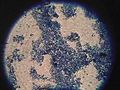
File:Crew6 MethBlue.jpg Meth blue microscope field of view(220 × 165 (19 KB)) - 17:07, 16 March 2020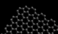
File:Graphene heptagon pentagon edge.png ...trates how the boundary of graphene is modified by a transmission electron microscope.(400 × 237 (39 KB)) - 02:41, 28 April 2013- ...phene sheet has been turned into a pattern of heptagons and pentagons by a microscope.]] ...raphene sheet is examined with a [[transmission electron microscope]], the microscope disrupts the edge and creates a boundary of alternating pentagonal and hept4 KB (549 words) - 13:55, 17 December 2018
- *Microscope *Microscope3 KB (364 words) - 08:36, 2 August 2019
- ...a mass of only 6 kg, the loaded equipment comprises of a regular camera, a microscope camera, a spectrometer, a subsurface radar and several ground penetrating t528 bytes (77 words) - 11:55, 17 December 2018
- Support Requested: Stereoscope and microscope. Support Requested: stereoscope and microscope7 KB (1,002 words) - 17:31, 7 March 2019
- ...d therefore form a more homogenous mass of small crystals (visible under a microscope) whereas the slow cooling rate of intrusive rocks allowed sufficient time f1 KB (226 words) - 08:52, 25 August 2021
- *optical microscope4 KB (572 words) - 00:36, 18 December 2018
- ...cientific community and the world media. Dr. McKay used scanning electron microscope (SEM) technology to image very fine slices of the meteorite. When he saw t9 KB (1,466 words) - 08:56, 25 August 2021
- with homemade microscopes in the event that a more official microscope is not available. ...re 1B). A pipette was used to transfer one drop of each soil sample onto a microscope slide. The field was examined at three resolutions (40x, 100x, and 1000x).42 KB (6,606 words) - 16:56, 7 March 2019
- ...ysis. We prepared some wet mount slides and analyzed the samples under the microscope in the science dome and found nothing unusual with regards cell structure. ...acterizing regolith samples and took photographs underneath the dissection microscope. A more thorough analysis of the samples will be available in tomorrow’s25 KB (3,795 words) - 17:40, 7 March 2019
- ...rom the GreenHab and wet mounts were prepared and viewed under the optical microscope. We found evidence of degraded cell structure, likely from the fungi growin8 KB (1,245 words) - 17:41, 7 March 2019
- ...rocess for the bacterial samples taken previously and attempted to use the microscope.9 KB (1,665 words) - 16:45, 7 March 2019
- ...on to the principal investigator for further analysis by scanning electron microscope, which we do not have access to here.14 KB (2,111 words) - 12:13, 24 March 2019
- ...utaraldehyde under refrigeration, a portion to be subjected to fluorescent microscope evaluation, and a portion to subject to the tertiary biology mission. In th ...en stop and spend several hours examining the contents of the foyer with a microscope. No, you give the place the once-over first. It is the same with field expl127 KB (20,888 words) - 21:33, 4 October 2019
- ...n the soil expanding and contracting due to major temperature changes. The microscope showed that the soil on top of the polygons is composed of flat particles (36 KB (5,518 words) - 14:50, 27 March 2024
- ...ed to the lab for further processing and inspection with an epifluorescent microscope. ...samples were inspected. Steve spent the several hours in the lab too. The microscope had been partially disassembled and Steve put the UV fluorescence parts bac118 KB (20,168 words) - 17:24, 16 March 2020
- ...d like more time to familiarize myself with this specific protocol and the microscope before attempting to draw definitive conclusions from the images. I am atta ...precludes biofilm formation. However, I think this is unlikely; under the microscope, the ‘Green’ element appeared to be some form of precipitate or salt, r157 KB (25,890 words) - 15:59, 5 November 2019
- ...that outlines the operation and maintenance of the biology lab, including microscope use and care, garbage, sterility issues, and sample preservation. This file ...ective samples were visualized using a bright field setting on the Olympus microscope with 200X-1000X magnification. In waypoint samples 5, 7, and 9, there were190 KB (31,918 words) - 09:28, 12 October 2019
- ...01 had microfossils inside of it. (See Image 8) Using a scanning electron microscope (SEM), McKay and his team imaged very fine slices of the meteorite. D. McKa64 KB (10,261 words) - 16:11, 21 December 2020